
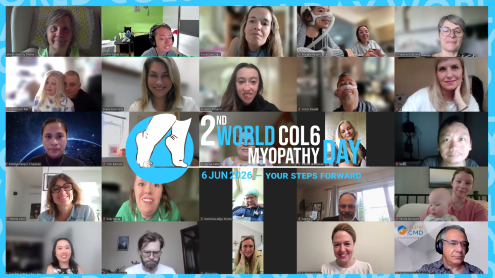
On June 6th, 2026, a very important milestone was reached: the second virtual edition of COL6 DAY. This achievement was made possible thanks to the outstanding commitment of many volunteers around the world and the dedicated members of several patient associations: Associazione Collagene VI Italia, Col6-Russia Association, CMD-Turkey, DGM-Muscular Dystrophy German Association, Fundación Noelia, the Ullrich Japanese Association, and the informal groups Col6-China, Col6-India, and Col6-LATAM.
We extend our heartfelt congratulations to everyone involved in organizing such a meaningful event, and we are grateful for the opportunity to take part in this important global moment in the journey of research and awareness for collagen VI-related myopathies and dystrophies.
This virtual congress brought together speakers and participants from all over the world, with more than 250 attendees across 8 panels and over 20 presentations. Representatives from across Europe, Asia, the United States, and Latin America took part. The event is increasingly becoming a global reference point for awareness, collaboration, and connection among patients, families, clinicians, and researchers working on COL6-related myopathies, their possible treatments, and their impact on daily life.
Among the topics presented were stem cell-based therapeutic approaches, gene therapy strategies to correct DNA defects, RNA-targeted therapies, epigenetic tools, mitochondrial-targeted treatments, collagen quantification methods, and increasingly refined cellular and tissue-engineered models for COL6 disorders.
Other presentations focused on patient management, diagnostic pathways, the importance of patient registries, population-based clinical data, orthopedic and respiratory care, and rehabilitation. Altogether, the congress highlighted the diversity and rapid development of the current research landscape in the field of COL6-associated myopathies.
Hidetoshi Sakurai, Japan
Topic: “Development of Cell Therapy for Collagen VI-Related Myopathies Using Mesenchymal Cells Derived from Induced Pluripotent Stem Cells (iPSCs)”
Professor Hidetoshi Sakurai presented promising work in regenerative medicine aimed at compensating for collagen VI deficiency by transplanting healthy cells capable of producing this protein.
In COL6-related myopathies such as Ullrich congenital muscular dystrophy and Bethlem myopathy, the lack of collagen VI disrupts muscle tissue structure, promotes fibrosis, and impairs muscle regeneration. Professor Sakurai’s group investigated whether this deficit could be compensated by injecting healthy mesenchymal stromal cells derived from induced pluripotent stem cells.
Using collagen VI knockout mice, the team showed that transplanted cells were able to restore collagen VI expression in muscle tissue, improve muscle regeneration after injury, reduce fibrosis, and increase muscle strength. The benefits were most pronounced when treatment was given early, especially from birth.
The group also compared different sources of mesenchymal stromal cells and found that iPSC-derived cells showed the best regenerative potential. Bone marrow-derived cells, by contrast, induced fibrosis in long-term observations. Based on these results, Professor Sakurai’s team is now working toward a first clinical study, tentatively projected for 2029. One of the major challenges remains reducing the risk of immune rejection after transplantation.
This work represents an important step toward a regenerative treatment strategy for COL6-related myopathies and offers cautious but real optimism for the future.
Numan Bulut, Turkey
Topic: “Assessment Methods for Patients with COL6-RD”
Numan Bulut presented a comprehensive overview of clinical assessment methods for people living with COL6-related disorders.
A complete evaluation should include neurological examination, family history, imaging such as MRI or ultrasound, respiratory function assessment, orthopedic and cardiac evaluation, and when needed, muscle biopsy. Physiotherapy assessments should be based on the full clinical picture and supported by standardized tools such as the Vignos scale, the Motor Function Measure, the six-minute walk test, balance testing, range of motion, and muscle strength measurements.
For non-ambulant patients, the longitudinal monitoring of contractures is particularly important. Respiratory follow-up was emphasized as one of the most critical prognostic factors, especially the decline in forced vital capacity, which may indicate the need to begin non-invasive ventilation.
The speaker also highlighted the value of the International Classification of Functioning, Disability and Health, which allows clinicians to assess not only motor ability but also participation, environmental factors, personal factors, pain, and fatigue. The main message was clear: assessment tools must be age-appropriate, disease-specific, reliable, and capable of tracking change over time.
Dr. Payam Mohassel, USA
Topic: “Regulation of TGF-β Activity in Skeletal Muscle Under the Influence of Collagen VI”
Dr. Payam Mohassel explored the relationship between collagen VI deficiency and the abnormal activation of TGF-β signaling in muscle.
Using collagen VI knockout mice, his group showed that the absence of collagen VI leads to marked muscle weakness, reduced contractile capacity, and fibrosis, including in the diaphragm. Transcriptomic analyses of patient biopsies revealed that disruption of the extracellular matrix is associated with abnormal activation of TGF-β, a signaling molecule that normally helps preserve tissue integrity but becomes harmful when chronically overactivated.
Because TGF-β signaling interacts with angiotensin II receptors, the team investigated whether losartan, a blood pressure medication, could reduce fibrosis by modulating this pathway. In a mouse study, three months of losartan treatment improved hanging performance, reduced fibrosis, and increased the number of healthy muscle nuclei, although body weight and chewing function did not change significantly.
These findings remain preliminary, but they point toward a potentially relevant antifibrotic strategy for COL6-related myopathies that deserves further study.
Dr. Malte Tiburcy, Germany
Topic: “Human Striated Muscle Tissue Models in COL6-Associated Dystrophy”
Dr. Malte Tiburcy presented an advanced tissue engineering platform designed to model COL6-related disease in human muscle.
Using patient-derived induced pluripotent stem cells, his group generates three-dimensional skeletal and cardiac muscle organoids in which cells produce their own extracellular matrix. These models contain not only muscle fibers but also satellite cells, neurons, and stromal cells such as fibroblasts, making them highly relevant for COL6 disorders.
The team created several mutated lines, including COL6A1 knockout models and lines carrying common COL6 mutations. These engineered tissues showed defective collagen VI assembly and impaired contractile performance under electrical stimulation, faithfully reproducing one of the key clinical features of COL6-related myopathies: muscle weakness.
The platform can grow up to 48 tissues in parallel and is suitable for drug screening. It was also used to model muscle injury and regeneration. Importantly, the group identified a distinct stromal cell population that emerged only under regenerative stress in the most severely affected mutant lines. This finding may help explain how chronic damage and atrophy contribute to disease progression.
This work provides a powerful preclinical tool for studying disease mechanisms and testing mutation-specific therapies.
Dr. Paolo Bernardi, Italy
Topic: “Development of New Treatments Targeting Mitochondrial Dysfunction in Collagen VI-Related Myopathies”
Professor Paolo Bernardi presented new therapeutic strategies aimed at correcting mitochondrial dysfunction, a key pathogenic mechanism in COL6-related myopathies.
His work focused on the mitochondrial permeability transition pore, whose abnormal opening contributes to calcium overload, mitochondrial depolarization, energy failure, and eventually muscle cell death. Although the primary defect in COL6 disease is extracellular, Professor Bernardi emphasized that altered calcium handling and mitochondrial dysfunction are central drivers of muscle weakness.
Through high-throughput screening and chemical optimization, his group developed a new diarylisoxazole compound that inhibits pore opening more effectively than previous agents and does so independently of cyclophilin D. The compound showed good stability, favorable pharmacological properties, and promising safety results.
In a mouse model of COL6-related myopathy, just four days of treatment led to measurable improvement: better mitochondrial calcium retention, reduced apoptosis, enhanced autophagy, improved muscle fiber structure, and increased muscle force. The speed of this response suggests that at least part of the weakness in COL6 disease may be functionally reversible.
If future studies confirm these findings, this approach could become one of the first targeted treatments aimed directly at a core disease mechanism in COL6-related myopathies.
Dr. Melania Giannotta, Italy
Topic: “Optimizing Surgical Outcomes: Focus on Perioperative Management in Patients with Collagen VI-Related Myopathies”
Dr. Melania Giannotta presented a highly relevant overview of perioperative management for patients undergoing orthopedic surgery, especially scoliosis correction.
Scoliosis in COL6-related myopathies can begin early, progress rapidly, and severely affect sitting balance, standing, walking, pain, and respiratory function. Respiratory decline often begins even before loss of ambulation, making pulmonary assessment and support essential parts of surgical planning.
The presentation reviewed indications for spinal surgery, including progressive curves greater than 40 degrees, worsening respiratory function, pain, and functional decline. Two main approaches were discussed: posterior spinal fusion for skeletally mature patients and growing constructs for younger children.
Special attention was given to anesthesia risks, including difficult airways, aspiration, cardiac evaluation, and the need to avoid succinylcholine because of the risk of rhabdomyolysis and hyperkalemia. Dr. Giannotta stressed the importance of multidisciplinary planning before, during, and after surgery, including respiratory preparation, nutritional optimization, physiotherapy, and postoperative mobilization.
A clinical case showed that with careful planning, spinal surgery can be both safe and effective in COL6 patients and may help stabilize respiratory decline over time.
Dr. Vittoria Cenni, Italy
Topic: “Morphological and Functional Changes in Tenocytes in Collagen VI-Related Myopathies”
Dr. Vittoria Cenni’s presentation highlighted the important but often underappreciated role of tendons in COL6-related disease.
Collagen VI is a key component not only of muscle extracellular matrix but also of tendon tissue. Her work showed that tendon involvement is not merely a secondary consequence of muscle weakness, but a primary effect of collagen VI deficiency.
In tendons from patients with Ullrich congenital muscular dystrophy, collagen fiber bundles were smaller, more disorganized, and structurally abnormal. Collagen VI failed to form a proper microfibrillar network and instead accumulated in aggregates. Similar abnormalities were observed in collagen I and collagen XII.
The study also revealed defects in mechanotransduction. Primary cilia were abnormally long, intracellular transport within them was altered, and signaling pathways such as Hedgehog and YAP responded poorly to mechanical stimulation. In addition, tenocytes from UCMD patients showed disorganized and non-directional migration in wound-healing assays.
These findings suggest that collagen VI deficiency disrupts how tendon cells sense and respond to mechanical cues, contributing directly to contractures and joint instability. They also reinforce the idea that future therapies must consider not only muscle but also connective tissues such as tendons.
Dr. Carsten Bönnemann, USA
Topic: “Gene Therapy Strategies for COL6-Associated Muscular Dystrophies”
Dr. Carsten Bönnemann delivered one of the most anticipated keynote lectures of the congress, offering a broad and clear overview of precision genetic medicine for COL6-related disorders.
He explained that COL6 disease can result from mutations in three genes, COL6A1, COL6A2, and COL6A3, and may follow either recessive or dominant mechanisms. This distinction is critical because the therapeutic strategy depends on the underlying genetic mechanism.
For recessive forms, one major approach is gene replacement using AAV vectors to deliver a healthy copy of the affected gene. However, the limited cargo capacity of AAV creates challenges, especially for COL6A3, which is too large to fit into a single vector.
For dominant forms, the mutant transcript must be selectively silenced or corrected. Strategies include antisense oligonucleotides and small interfering RNAs that target the abnormal messenger RNA without affecting the healthy allele. In patient-derived fibroblasts, silencing the mutant transcript has already restored collagen VI secretion and extracellular matrix assembly.
Dr. Bönnemann also discussed genome editing technologies such as CRISPR-Cas9, base editing, and prime editing. These approaches offer the possibility of permanent correction, but they still face major challenges, especially delivery to the right target cells. In COL6 disease, that key target is not only the muscle fiber itself, but especially the fibroblast, the cell type primarily responsible for producing collagen VI.
The message of the talk was encouraging: while the biological and technical challenges are real, precision genetic medicine for COL6 disorders is no longer theoretical. It is actively being developed.
Dr. Valérie Allamand, France
Topic: “Skin Fibroblasts as a Model for COL6 Myopathies and Therapeutic Approaches”
Dr. Valérie Allamand presented elegant work using skin fibroblasts as an accessible and informative model for COL6 disease.
Because fibroblasts actively produce collagen VI, they allow researchers to assess secretion defects and extracellular matrix organization. Her team has developed an optimized culture protocol called OptiMEC, which increases the production of extracellular matrix components and makes disease-related abnormalities more visible.
Under these conditions, fibroblasts from patients with heterozygous mutations showed increased collagen VI secretion and improved structural organization of the matrix, allowing more subtle defects to be detected more reliably.
In collaboration with Dr. Bönnemann’s group, the team also tested antisense oligonucleotides designed to suppress mutant COL6A3 transcripts associated with exon skipping. Under OptiMEC conditions, the treatment significantly improved collagen VI secretion and extracellular network formation.
The laboratory is also developing RNA-based approaches for homozygous mutations, although these remain at an early stage. Altogether, the work provides both a strong disease model and an experimental platform for evaluating future therapies.
Dr. Virginia Arechavala-Gómez, Spain
Topic: “Optimizing Diagnosis of COL6-Associated Dystrophies Through Collablot Analysis”
Dr. Virginia Arechavala-Gómez presented Collablot, a semi-quantitative platform designed to improve the diagnosis of COL6-related disorders.
Based on western blot-like principles, the method evaluates both how much collagen VI is secreted into the extracellular matrix and how well it is organized into fibrillar networks. This makes it possible to assess both quantity and quality at the same time.
When applied to larger collections of patient fibroblasts, including complex or uncertain cases, the method proved useful for identifying collagen VI pathology even when a causative mutation had not yet been clearly established.
Collablot is also being used to assess therapeutic effects, including restoration of correct splicing after gene editing or RNA-based intervention. Because of its relative simplicity and scalability, it could become an important support tool in routine diagnostic practice.
Dr. Jessica Moraes, United Kingdom
Topic: “The Global COL6-RD Registry – John Walton Muscular Dystrophy Research Centre”
Dr. Jessica Moraes presented the global COL6-RD registry, an essential resource for future clinical trials and natural history studies.
The registry is an online platform where patients or their families can enter clinical and genetic information, which is then verified by clinicians and researchers. It collects data such as age, country of residence, genotype, phenotype, functional status, symptoms, and treatments.
At present, the registry includes patients from 57 countries, with the largest numbers coming from the United States and the United Kingdom, followed by Germany, France, and Italy. The sex distribution is approximately equal, and the largest genetic subgroup includes mutations in COL6A1.
The registry plays a central role in connecting patients with research, defining the natural history of the disease, improving standards of care, and preparing for future international clinical trials. Dr. Moraes encouraged broader global participation to strengthen its impact.
Dr. Yunsong Jiang, United Kingdom
Topic: “New Human 3D Muscle Models with Extracellular Matrix for the Study of COL6 Muscular Dystrophies and the Search for Treatments”
Dr. Yunsong Jiang presented an innovative 3D human muscle platform developed in the laboratory of Professor Francesco Saverio Tedesco.
These miniature engineered muscles combine muscle cells with fibroblasts, allowing researchers to recreate a much more realistic extracellular matrix environment than standard two-dimensional cultures. When healthy muscle cells were combined with fibroblasts from UCMD or Bethlem patients, the tissues developed striking macroscopic changes resembling contractures.
The diseased models also showed fragmented and disorganized collagen VI networks, increased TGF-β signaling, altered extracellular matrix remodeling, and markedly impaired contractility under electrical stimulation.
This platform is the first to reproduce multiple key features of COL6-related myopathies in a human 3D system, from structural matrix defects to functional muscle weakness. It is expected to play an important role in therapeutic testing.
Dr. Reghan Foley, USA
Topic: “The Spectrum of COL6-Related Myopathies – From Natural History to Clinical Trial Readiness”
Dr. Reghan Foley presented a comprehensive overview of the clinical spectrum of COL6-related myopathies and the current efforts to prepare the field for clinical trials.
She reviewed the three main phenotypes — Ullrich congenital muscular dystrophy, intermediate forms, and Bethlem myopathy — and explained how early motor milestones can help predict disease course. She also emphasized the importance of respiratory function as a key prognostic marker.
In Ullrich patients, forced vital capacity declines by about 2.6% per year, with the need for non-invasive ventilation typically arising around age 11. In the intermediate group, respiratory decline is slower and NIV often begins around age 21. In Bethlem myopathy, respiratory support is less common and usually needed much later in adulthood.
Dr. Foley also discussed a deep intronic COL6A1 variant that defines a particularly important subgroup for RNA-targeted therapy. A mosaic patient carrying this variant showed a much milder phenotype, providing a real-life example of how partial correction of a mutant transcript could shift disease severity.
She concluded by outlining the three essential pillars of clinical trial readiness: detailed natural history studies, robust and meaningful outcome measures, and optimized clinical care. She also reviewed skin manifestations, hair loss, and ongoing biomarker studies using blood and urine.
Dr. Claire Baldock, United Kingdom
Topic: “Unraveling the Structure of Collagen VI Microfibrils by Cryo-Electron Microscopy”
Professor Claire Baldock presented groundbreaking structural work on collagen VI microfibrils using cryo-electron microscopy.
Collagen VI is a non-fibrillar collagen with a highly specialized assembly process. Her team developed systems to study both recombinant mini-collagen VI proteins and native microfibrils isolated from tissues. This allowed them to investigate how the three alpha chains assemble into stable heterotrimers and then into larger extracellular microfibrils.
For the first time, the group obtained a high-resolution structure of native collagen VI microfibrils. They showed how the alpha chains are arranged, how von Willebrand factor A domains are positioned, and how adjacent tetramers interact to form the final structure.
These findings are highly relevant for the COL6 field because they make it possible to map pathogenic variants more precisely and understand how specific mutations disrupt collagen VI assembly. This is a major step forward for molecular diagnosis and the design of targeted therapies.
Dr. Veronique Bolduc, USA
Topic: “Development of a Targeted RNA Therapy for the COL6A1 c.930+189C>T Variant”
Dr. Veronique Bolduc presented a highly precise example of personalized medicine: an RNA-targeted therapy developed specifically for the deep intronic COL6A1 c.930+189C>T variant.
Her team used antisense oligonucleotides to block the use of the pathological splice fragment. In patient cells, this sharply reduced the mutant RNA, lowered the level of abnormal protein, and restored the collagen VI network to near-normal levels.
To test the therapy in vivo, the researchers created a humanized mouse model carrying the mutation. These mice developed muscle weakness and pathological changes consistent with muscular dystrophy, confirming that the model is suitable for preclinical studies.
The group has already shown in vivo delivery to muscle and specific cell types. They are also exploring alternative approaches, including U7-mediated splicing modulation via AAV vectors and base editing. One of the key remaining challenges is efficient delivery to fibro-adipogenic progenitors, but active work is ongoing.
This strategy is designed for a single variant, but once the platform is refined, it could potentially be adapted more quickly to other mutations of the same class.
Sergey A. Kurbatov, Russia
Topic: “Muscle MRI and Needle Electromyography in the Diagnosis of Collagen VI-Related Myopathies”
Sergey A. Kurbatov discussed the role of muscle MRI and needle EMG in the diagnosis of COL6-related myopathies, with a special focus on diagnostic pitfalls.
He emphasized that although muscle MRI patterns such as the well-known “tigroid” appearance are often associated with COL6 disease, they are not completely specific and can also appear in other muscular dystrophies. Likewise, EMG may show neurogenic-like patterns in genetically confirmed COL6 patients, especially in recessive COL6A3-related cases, which can lead to confusion with spinal muscular atrophy.
A particularly important example involved the COL6A3 c.7447A>G variant, which was repeatedly associated with pseudo-neurogenic presentations and delayed diagnosis. The main clinical takeaway was that diagnosis should always rely on integrated clinical, imaging, electrophysiological, and molecular evaluation.
Diana S. Abramova, Russia
Topic: “The Russian COL6-RD Patient Registry: Clinical-Genetic Profile and Preliminary Results from the First Two Years of Follow-Up”
Diana S. Abramova presented data from the Russian COL6-RD patient registry, established in 2024 through collaboration between the patient and medical communities.
In just two years, the registry has grown to 203 patients, 197 of whom have genetically confirmed diagnoses. Around 70% of patients entered through the patient community itself, underlining the vital role of family awareness and advocacy.
The largest subgroup carries mutations in COL6A1, while COL6A3 shows the widest mutational diversity. Particularly notable is the c.7447A>G variant in COL6A3, for which the Russian cohort appears to be the largest national cohort reported so far.
The registry has already revealed important trends in ambulation loss, respiratory decline, and orthopedic complications, and it is becoming a critical foundation for future natural history studies and therapeutic readiness.
Dmitry A. Feklistov, Russia
Topic: “Orthopedic Management of Russian Patients with COL6-RD: Clinical Experience”
Dmitry A. Feklistov summarized twenty years of orthopedic experience in the management of neuromuscular patients, with a focus on contractures, scoliosis, and hip instability in COL6-related disorders.
He stressed that timely orthopedic care is becoming even more important as disease-modifying therapies approach the clinic. If contractures and spinal deformities are not managed early, they may significantly reduce the benefit of future treatments.
The presentation reviewed conservative measures such as ankle-foot orthoses, knee-ankle-foot orthoses, wrist splints, and spinal braces, while also acknowledging the limited disease-specific evidence supporting these approaches. Surgery remains the most reliable method for correcting severe scoliosis and can significantly improve quality of life, posture, and respiratory function.
The central message was that the field urgently needs international, standardized orthopedic care protocols specifically adapted to COL6-related myopathies.
Evgenia A. Melnik, Russia
Topic: “COL12A1-Related Myopathies: Expanding the Differential Diagnosis of COL6-Related Disorders”
Evgenia A. Melnik presented an important talk on COL12A1-related myopathies, which can clinically resemble COL6-related disease.
COL12A1 encodes collagen XII, another extracellular matrix protein involved in the response of connective tissue to mechanical stress. Mutations in this gene can lead to phenotypes overlapping with Ullrich and Bethlem-like disorders.
The presentation reviewed the clinical spectrum of COL12A1-related myopathy, including recessive severe early-onset forms and dominant forms with contractures, scoliosis, distal weakness, and sometimes overlap with hypermobility syndromes. MRI patterns differ from classic COL6 disease and may involve abdominal and neck muscles.
The main practical conclusion was that COL12A1 should be included in the differential diagnosis when COL6 testing is negative but the phenotype remains strongly suggestive of a collagen-related myopathy.
Alexander R. Stevanovic, Russia
Topic: “Rehabilitation and Selection of Technical Aids for Patients with COL6-RD: Clinical Aspects”
Alexander R. Stevanovic concluded with a practical and patient-centered presentation on rehabilitation.
He emphasized a core principle: low intensity, high regularity. Rehabilitation in COL6-related myopathies must support function without overloading already fragile and easily fatigued muscles. The goal is always maximum function with minimum pathology.
He described a “24/7” approach that includes medication when prescribed, individualized physiotherapy, proper positioning throughout the day, orthotic support, and surgery when appropriate. Daily stretching, hydrotherapy, respiratory exercises, occupational therapy, and environmental adaptations all play important roles.
Special emphasis was placed on positioning and standing. Proper seating helps prevent asymmetry and scoliosis, while supported standing can reduce the risk of hip displacement, preserve bone density, and improve the function of multiple organ systems. The overall message was powerful: until truly curative therapies become available, structured rehabilitation remains one of the most important ways to preserve health and quality of life.
Conclusion
Whether presented by scientists, clinicians, therapists, patient advocates, or families, every contribution helped build a clearer and more hopeful future for the COL6 community. This second COL6 DAY Virtual Congress once again showed how much progress is being made across multiple areas: gene and RNA therapies, regenerative medicine, mitochondrial biology, diagnostics, clinical care, rehabilitation, and patient registries.
Above all, the congress reaffirmed something essential: meaningful progress in rare diseases happens when patients, families, researchers, and clinicians work together across borders.
This global awareness day is more than a symbolic occasion. It is a space to learn, to connect, to share knowledge, and to strengthen the international network that our community needs. Because we are rare, we must think globally in order to be stronger together.
We hope this second edition will be followed by many more, and we warmly invite everyone to continue taking part in this journey with us.
We extend our heartfelt congratulations to everyone involved in organizing such a meaningful event, and we are grateful for the opportunity to take part in this important global moment in the journey of research and awareness for collagen VI-related myopathies and dystrophies.
This virtual congress brought together speakers and participants from all over the world, with more than 250 attendees across 8 panels and over 20 presentations. Representatives from across Europe, Asia, the United States, and Latin America took part. The event is increasingly becoming a global reference point for awareness, collaboration, and connection among patients, families, clinicians, and researchers working on COL6-related myopathies, their possible treatments, and their impact on daily life.
Among the topics presented were stem cell-based therapeutic approaches, gene therapy strategies to correct DNA defects, RNA-targeted therapies, epigenetic tools, mitochondrial-targeted treatments, collagen quantification methods, and increasingly refined cellular and tissue-engineered models for COL6 disorders.
Other presentations focused on patient management, diagnostic pathways, the importance of patient registries, population-based clinical data, orthopedic and respiratory care, and rehabilitation. Altogether, the congress highlighted the diversity and rapid development of the current research landscape in the field of COL6-associated myopathies.
Hidetoshi Sakurai, Japan
Topic: “Development of Cell Therapy for Collagen VI-Related Myopathies Using Mesenchymal Cells Derived from Induced Pluripotent Stem Cells (iPSCs)”
Professor Hidetoshi Sakurai presented promising work in regenerative medicine aimed at compensating for collagen VI deficiency by transplanting healthy cells capable of producing this protein.
In COL6-related myopathies such as Ullrich congenital muscular dystrophy and Bethlem myopathy, the lack of collagen VI disrupts muscle tissue structure, promotes fibrosis, and impairs muscle regeneration. Professor Sakurai’s group investigated whether this deficit could be compensated by injecting healthy mesenchymal stromal cells derived from induced pluripotent stem cells.
Using collagen VI knockout mice, the team showed that transplanted cells were able to restore collagen VI expression in muscle tissue, improve muscle regeneration after injury, reduce fibrosis, and increase muscle strength. The benefits were most pronounced when treatment was given early, especially from birth.
The group also compared different sources of mesenchymal stromal cells and found that iPSC-derived cells showed the best regenerative potential. Bone marrow-derived cells, by contrast, induced fibrosis in long-term observations. Based on these results, Professor Sakurai’s team is now working toward a first clinical study, tentatively projected for 2029. One of the major challenges remains reducing the risk of immune rejection after transplantation.
This work represents an important step toward a regenerative treatment strategy for COL6-related myopathies and offers cautious but real optimism for the future.
Numan Bulut, Turkey
Topic: “Assessment Methods for Patients with COL6-RD”
Numan Bulut presented a comprehensive overview of clinical assessment methods for people living with COL6-related disorders.
A complete evaluation should include neurological examination, family history, imaging such as MRI or ultrasound, respiratory function assessment, orthopedic and cardiac evaluation, and when needed, muscle biopsy. Physiotherapy assessments should be based on the full clinical picture and supported by standardized tools such as the Vignos scale, the Motor Function Measure, the six-minute walk test, balance testing, range of motion, and muscle strength measurements.
For non-ambulant patients, the longitudinal monitoring of contractures is particularly important. Respiratory follow-up was emphasized as one of the most critical prognostic factors, especially the decline in forced vital capacity, which may indicate the need to begin non-invasive ventilation.
The speaker also highlighted the value of the International Classification of Functioning, Disability and Health, which allows clinicians to assess not only motor ability but also participation, environmental factors, personal factors, pain, and fatigue. The main message was clear: assessment tools must be age-appropriate, disease-specific, reliable, and capable of tracking change over time.
Dr. Payam Mohassel, USA
Topic: “Regulation of TGF-β Activity in Skeletal Muscle Under the Influence of Collagen VI”
Dr. Payam Mohassel explored the relationship between collagen VI deficiency and the abnormal activation of TGF-β signaling in muscle.
Using collagen VI knockout mice, his group showed that the absence of collagen VI leads to marked muscle weakness, reduced contractile capacity, and fibrosis, including in the diaphragm. Transcriptomic analyses of patient biopsies revealed that disruption of the extracellular matrix is associated with abnormal activation of TGF-β, a signaling molecule that normally helps preserve tissue integrity but becomes harmful when chronically overactivated.
Because TGF-β signaling interacts with angiotensin II receptors, the team investigated whether losartan, a blood pressure medication, could reduce fibrosis by modulating this pathway. In a mouse study, three months of losartan treatment improved hanging performance, reduced fibrosis, and increased the number of healthy muscle nuclei, although body weight and chewing function did not change significantly.
These findings remain preliminary, but they point toward a potentially relevant antifibrotic strategy for COL6-related myopathies that deserves further study.
Dr. Malte Tiburcy, Germany
Topic: “Human Striated Muscle Tissue Models in COL6-Associated Dystrophy”
Dr. Malte Tiburcy presented an advanced tissue engineering platform designed to model COL6-related disease in human muscle.
Using patient-derived induced pluripotent stem cells, his group generates three-dimensional skeletal and cardiac muscle organoids in which cells produce their own extracellular matrix. These models contain not only muscle fibers but also satellite cells, neurons, and stromal cells such as fibroblasts, making them highly relevant for COL6 disorders.
The team created several mutated lines, including COL6A1 knockout models and lines carrying common COL6 mutations. These engineered tissues showed defective collagen VI assembly and impaired contractile performance under electrical stimulation, faithfully reproducing one of the key clinical features of COL6-related myopathies: muscle weakness.
The platform can grow up to 48 tissues in parallel and is suitable for drug screening. It was also used to model muscle injury and regeneration. Importantly, the group identified a distinct stromal cell population that emerged only under regenerative stress in the most severely affected mutant lines. This finding may help explain how chronic damage and atrophy contribute to disease progression.
This work provides a powerful preclinical tool for studying disease mechanisms and testing mutation-specific therapies.
Dr. Paolo Bernardi, Italy
Topic: “Development of New Treatments Targeting Mitochondrial Dysfunction in Collagen VI-Related Myopathies”
Professor Paolo Bernardi presented new therapeutic strategies aimed at correcting mitochondrial dysfunction, a key pathogenic mechanism in COL6-related myopathies.
His work focused on the mitochondrial permeability transition pore, whose abnormal opening contributes to calcium overload, mitochondrial depolarization, energy failure, and eventually muscle cell death. Although the primary defect in COL6 disease is extracellular, Professor Bernardi emphasized that altered calcium handling and mitochondrial dysfunction are central drivers of muscle weakness.
Through high-throughput screening and chemical optimization, his group developed a new diarylisoxazole compound that inhibits pore opening more effectively than previous agents and does so independently of cyclophilin D. The compound showed good stability, favorable pharmacological properties, and promising safety results.
In a mouse model of COL6-related myopathy, just four days of treatment led to measurable improvement: better mitochondrial calcium retention, reduced apoptosis, enhanced autophagy, improved muscle fiber structure, and increased muscle force. The speed of this response suggests that at least part of the weakness in COL6 disease may be functionally reversible.
If future studies confirm these findings, this approach could become one of the first targeted treatments aimed directly at a core disease mechanism in COL6-related myopathies.
Dr. Melania Giannotta, Italy
Topic: “Optimizing Surgical Outcomes: Focus on Perioperative Management in Patients with Collagen VI-Related Myopathies”
Dr. Melania Giannotta presented a highly relevant overview of perioperative management for patients undergoing orthopedic surgery, especially scoliosis correction.
Scoliosis in COL6-related myopathies can begin early, progress rapidly, and severely affect sitting balance, standing, walking, pain, and respiratory function. Respiratory decline often begins even before loss of ambulation, making pulmonary assessment and support essential parts of surgical planning.
The presentation reviewed indications for spinal surgery, including progressive curves greater than 40 degrees, worsening respiratory function, pain, and functional decline. Two main approaches were discussed: posterior spinal fusion for skeletally mature patients and growing constructs for younger children.
Special attention was given to anesthesia risks, including difficult airways, aspiration, cardiac evaluation, and the need to avoid succinylcholine because of the risk of rhabdomyolysis and hyperkalemia. Dr. Giannotta stressed the importance of multidisciplinary planning before, during, and after surgery, including respiratory preparation, nutritional optimization, physiotherapy, and postoperative mobilization.
A clinical case showed that with careful planning, spinal surgery can be both safe and effective in COL6 patients and may help stabilize respiratory decline over time.
Dr. Vittoria Cenni, Italy
Topic: “Morphological and Functional Changes in Tenocytes in Collagen VI-Related Myopathies”
Dr. Vittoria Cenni’s presentation highlighted the important but often underappreciated role of tendons in COL6-related disease.
Collagen VI is a key component not only of muscle extracellular matrix but also of tendon tissue. Her work showed that tendon involvement is not merely a secondary consequence of muscle weakness, but a primary effect of collagen VI deficiency.
In tendons from patients with Ullrich congenital muscular dystrophy, collagen fiber bundles were smaller, more disorganized, and structurally abnormal. Collagen VI failed to form a proper microfibrillar network and instead accumulated in aggregates. Similar abnormalities were observed in collagen I and collagen XII.
The study also revealed defects in mechanotransduction. Primary cilia were abnormally long, intracellular transport within them was altered, and signaling pathways such as Hedgehog and YAP responded poorly to mechanical stimulation. In addition, tenocytes from UCMD patients showed disorganized and non-directional migration in wound-healing assays.
These findings suggest that collagen VI deficiency disrupts how tendon cells sense and respond to mechanical cues, contributing directly to contractures and joint instability. They also reinforce the idea that future therapies must consider not only muscle but also connective tissues such as tendons.
Dr. Carsten Bönnemann, USA
Topic: “Gene Therapy Strategies for COL6-Associated Muscular Dystrophies”
Dr. Carsten Bönnemann delivered one of the most anticipated keynote lectures of the congress, offering a broad and clear overview of precision genetic medicine for COL6-related disorders.
He explained that COL6 disease can result from mutations in three genes, COL6A1, COL6A2, and COL6A3, and may follow either recessive or dominant mechanisms. This distinction is critical because the therapeutic strategy depends on the underlying genetic mechanism.
For recessive forms, one major approach is gene replacement using AAV vectors to deliver a healthy copy of the affected gene. However, the limited cargo capacity of AAV creates challenges, especially for COL6A3, which is too large to fit into a single vector.
For dominant forms, the mutant transcript must be selectively silenced or corrected. Strategies include antisense oligonucleotides and small interfering RNAs that target the abnormal messenger RNA without affecting the healthy allele. In patient-derived fibroblasts, silencing the mutant transcript has already restored collagen VI secretion and extracellular matrix assembly.
Dr. Bönnemann also discussed genome editing technologies such as CRISPR-Cas9, base editing, and prime editing. These approaches offer the possibility of permanent correction, but they still face major challenges, especially delivery to the right target cells. In COL6 disease, that key target is not only the muscle fiber itself, but especially the fibroblast, the cell type primarily responsible for producing collagen VI.
The message of the talk was encouraging: while the biological and technical challenges are real, precision genetic medicine for COL6 disorders is no longer theoretical. It is actively being developed.
Dr. Valérie Allamand, France
Topic: “Skin Fibroblasts as a Model for COL6 Myopathies and Therapeutic Approaches”
Dr. Valérie Allamand presented elegant work using skin fibroblasts as an accessible and informative model for COL6 disease.
Because fibroblasts actively produce collagen VI, they allow researchers to assess secretion defects and extracellular matrix organization. Her team has developed an optimized culture protocol called OptiMEC, which increases the production of extracellular matrix components and makes disease-related abnormalities more visible.
Under these conditions, fibroblasts from patients with heterozygous mutations showed increased collagen VI secretion and improved structural organization of the matrix, allowing more subtle defects to be detected more reliably.
In collaboration with Dr. Bönnemann’s group, the team also tested antisense oligonucleotides designed to suppress mutant COL6A3 transcripts associated with exon skipping. Under OptiMEC conditions, the treatment significantly improved collagen VI secretion and extracellular network formation.
The laboratory is also developing RNA-based approaches for homozygous mutations, although these remain at an early stage. Altogether, the work provides both a strong disease model and an experimental platform for evaluating future therapies.
Dr. Virginia Arechavala-Gómez, Spain
Topic: “Optimizing Diagnosis of COL6-Associated Dystrophies Through Collablot Analysis”
Dr. Virginia Arechavala-Gómez presented Collablot, a semi-quantitative platform designed to improve the diagnosis of COL6-related disorders.
Based on western blot-like principles, the method evaluates both how much collagen VI is secreted into the extracellular matrix and how well it is organized into fibrillar networks. This makes it possible to assess both quantity and quality at the same time.
When applied to larger collections of patient fibroblasts, including complex or uncertain cases, the method proved useful for identifying collagen VI pathology even when a causative mutation had not yet been clearly established.
Collablot is also being used to assess therapeutic effects, including restoration of correct splicing after gene editing or RNA-based intervention. Because of its relative simplicity and scalability, it could become an important support tool in routine diagnostic practice.
Dr. Jessica Moraes, United Kingdom
Topic: “The Global COL6-RD Registry – John Walton Muscular Dystrophy Research Centre”
Dr. Jessica Moraes presented the global COL6-RD registry, an essential resource for future clinical trials and natural history studies.
The registry is an online platform where patients or their families can enter clinical and genetic information, which is then verified by clinicians and researchers. It collects data such as age, country of residence, genotype, phenotype, functional status, symptoms, and treatments.
At present, the registry includes patients from 57 countries, with the largest numbers coming from the United States and the United Kingdom, followed by Germany, France, and Italy. The sex distribution is approximately equal, and the largest genetic subgroup includes mutations in COL6A1.
The registry plays a central role in connecting patients with research, defining the natural history of the disease, improving standards of care, and preparing for future international clinical trials. Dr. Moraes encouraged broader global participation to strengthen its impact.
Dr. Yunsong Jiang, United Kingdom
Topic: “New Human 3D Muscle Models with Extracellular Matrix for the Study of COL6 Muscular Dystrophies and the Search for Treatments”
Dr. Yunsong Jiang presented an innovative 3D human muscle platform developed in the laboratory of Professor Francesco Saverio Tedesco.
These miniature engineered muscles combine muscle cells with fibroblasts, allowing researchers to recreate a much more realistic extracellular matrix environment than standard two-dimensional cultures. When healthy muscle cells were combined with fibroblasts from UCMD or Bethlem patients, the tissues developed striking macroscopic changes resembling contractures.
The diseased models also showed fragmented and disorganized collagen VI networks, increased TGF-β signaling, altered extracellular matrix remodeling, and markedly impaired contractility under electrical stimulation.
This platform is the first to reproduce multiple key features of COL6-related myopathies in a human 3D system, from structural matrix defects to functional muscle weakness. It is expected to play an important role in therapeutic testing.
Dr. Reghan Foley, USA
Topic: “The Spectrum of COL6-Related Myopathies – From Natural History to Clinical Trial Readiness”
Dr. Reghan Foley presented a comprehensive overview of the clinical spectrum of COL6-related myopathies and the current efforts to prepare the field for clinical trials.
She reviewed the three main phenotypes — Ullrich congenital muscular dystrophy, intermediate forms, and Bethlem myopathy — and explained how early motor milestones can help predict disease course. She also emphasized the importance of respiratory function as a key prognostic marker.
In Ullrich patients, forced vital capacity declines by about 2.6% per year, with the need for non-invasive ventilation typically arising around age 11. In the intermediate group, respiratory decline is slower and NIV often begins around age 21. In Bethlem myopathy, respiratory support is less common and usually needed much later in adulthood.
Dr. Foley also discussed a deep intronic COL6A1 variant that defines a particularly important subgroup for RNA-targeted therapy. A mosaic patient carrying this variant showed a much milder phenotype, providing a real-life example of how partial correction of a mutant transcript could shift disease severity.
She concluded by outlining the three essential pillars of clinical trial readiness: detailed natural history studies, robust and meaningful outcome measures, and optimized clinical care. She also reviewed skin manifestations, hair loss, and ongoing biomarker studies using blood and urine.
Dr. Claire Baldock, United Kingdom
Topic: “Unraveling the Structure of Collagen VI Microfibrils by Cryo-Electron Microscopy”
Professor Claire Baldock presented groundbreaking structural work on collagen VI microfibrils using cryo-electron microscopy.
Collagen VI is a non-fibrillar collagen with a highly specialized assembly process. Her team developed systems to study both recombinant mini-collagen VI proteins and native microfibrils isolated from tissues. This allowed them to investigate how the three alpha chains assemble into stable heterotrimers and then into larger extracellular microfibrils.
For the first time, the group obtained a high-resolution structure of native collagen VI microfibrils. They showed how the alpha chains are arranged, how von Willebrand factor A domains are positioned, and how adjacent tetramers interact to form the final structure.
These findings are highly relevant for the COL6 field because they make it possible to map pathogenic variants more precisely and understand how specific mutations disrupt collagen VI assembly. This is a major step forward for molecular diagnosis and the design of targeted therapies.
Dr. Veronique Bolduc, USA
Topic: “Development of a Targeted RNA Therapy for the COL6A1 c.930+189C>T Variant”
Dr. Veronique Bolduc presented a highly precise example of personalized medicine: an RNA-targeted therapy developed specifically for the deep intronic COL6A1 c.930+189C>T variant.
Her team used antisense oligonucleotides to block the use of the pathological splice fragment. In patient cells, this sharply reduced the mutant RNA, lowered the level of abnormal protein, and restored the collagen VI network to near-normal levels.
To test the therapy in vivo, the researchers created a humanized mouse model carrying the mutation. These mice developed muscle weakness and pathological changes consistent with muscular dystrophy, confirming that the model is suitable for preclinical studies.
The group has already shown in vivo delivery to muscle and specific cell types. They are also exploring alternative approaches, including U7-mediated splicing modulation via AAV vectors and base editing. One of the key remaining challenges is efficient delivery to fibro-adipogenic progenitors, but active work is ongoing.
This strategy is designed for a single variant, but once the platform is refined, it could potentially be adapted more quickly to other mutations of the same class.
Sergey A. Kurbatov, Russia
Topic: “Muscle MRI and Needle Electromyography in the Diagnosis of Collagen VI-Related Myopathies”
Sergey A. Kurbatov discussed the role of muscle MRI and needle EMG in the diagnosis of COL6-related myopathies, with a special focus on diagnostic pitfalls.
He emphasized that although muscle MRI patterns such as the well-known “tigroid” appearance are often associated with COL6 disease, they are not completely specific and can also appear in other muscular dystrophies. Likewise, EMG may show neurogenic-like patterns in genetically confirmed COL6 patients, especially in recessive COL6A3-related cases, which can lead to confusion with spinal muscular atrophy.
A particularly important example involved the COL6A3 c.7447A>G variant, which was repeatedly associated with pseudo-neurogenic presentations and delayed diagnosis. The main clinical takeaway was that diagnosis should always rely on integrated clinical, imaging, electrophysiological, and molecular evaluation.
Diana S. Abramova, Russia
Topic: “The Russian COL6-RD Patient Registry: Clinical-Genetic Profile and Preliminary Results from the First Two Years of Follow-Up”
Diana S. Abramova presented data from the Russian COL6-RD patient registry, established in 2024 through collaboration between the patient and medical communities.
In just two years, the registry has grown to 203 patients, 197 of whom have genetically confirmed diagnoses. Around 70% of patients entered through the patient community itself, underlining the vital role of family awareness and advocacy.
The largest subgroup carries mutations in COL6A1, while COL6A3 shows the widest mutational diversity. Particularly notable is the c.7447A>G variant in COL6A3, for which the Russian cohort appears to be the largest national cohort reported so far.
The registry has already revealed important trends in ambulation loss, respiratory decline, and orthopedic complications, and it is becoming a critical foundation for future natural history studies and therapeutic readiness.
Dmitry A. Feklistov, Russia
Topic: “Orthopedic Management of Russian Patients with COL6-RD: Clinical Experience”
Dmitry A. Feklistov summarized twenty years of orthopedic experience in the management of neuromuscular patients, with a focus on contractures, scoliosis, and hip instability in COL6-related disorders.
He stressed that timely orthopedic care is becoming even more important as disease-modifying therapies approach the clinic. If contractures and spinal deformities are not managed early, they may significantly reduce the benefit of future treatments.
The presentation reviewed conservative measures such as ankle-foot orthoses, knee-ankle-foot orthoses, wrist splints, and spinal braces, while also acknowledging the limited disease-specific evidence supporting these approaches. Surgery remains the most reliable method for correcting severe scoliosis and can significantly improve quality of life, posture, and respiratory function.
The central message was that the field urgently needs international, standardized orthopedic care protocols specifically adapted to COL6-related myopathies.
Evgenia A. Melnik, Russia
Topic: “COL12A1-Related Myopathies: Expanding the Differential Diagnosis of COL6-Related Disorders”
Evgenia A. Melnik presented an important talk on COL12A1-related myopathies, which can clinically resemble COL6-related disease.
COL12A1 encodes collagen XII, another extracellular matrix protein involved in the response of connective tissue to mechanical stress. Mutations in this gene can lead to phenotypes overlapping with Ullrich and Bethlem-like disorders.
The presentation reviewed the clinical spectrum of COL12A1-related myopathy, including recessive severe early-onset forms and dominant forms with contractures, scoliosis, distal weakness, and sometimes overlap with hypermobility syndromes. MRI patterns differ from classic COL6 disease and may involve abdominal and neck muscles.
The main practical conclusion was that COL12A1 should be included in the differential diagnosis when COL6 testing is negative but the phenotype remains strongly suggestive of a collagen-related myopathy.
Alexander R. Stevanovic, Russia
Topic: “Rehabilitation and Selection of Technical Aids for Patients with COL6-RD: Clinical Aspects”
Alexander R. Stevanovic concluded with a practical and patient-centered presentation on rehabilitation.
He emphasized a core principle: low intensity, high regularity. Rehabilitation in COL6-related myopathies must support function without overloading already fragile and easily fatigued muscles. The goal is always maximum function with minimum pathology.
He described a “24/7” approach that includes medication when prescribed, individualized physiotherapy, proper positioning throughout the day, orthotic support, and surgery when appropriate. Daily stretching, hydrotherapy, respiratory exercises, occupational therapy, and environmental adaptations all play important roles.
Special emphasis was placed on positioning and standing. Proper seating helps prevent asymmetry and scoliosis, while supported standing can reduce the risk of hip displacement, preserve bone density, and improve the function of multiple organ systems. The overall message was powerful: until truly curative therapies become available, structured rehabilitation remains one of the most important ways to preserve health and quality of life.
Conclusion
Whether presented by scientists, clinicians, therapists, patient advocates, or families, every contribution helped build a clearer and more hopeful future for the COL6 community. This second COL6 DAY Virtual Congress once again showed how much progress is being made across multiple areas: gene and RNA therapies, regenerative medicine, mitochondrial biology, diagnostics, clinical care, rehabilitation, and patient registries.
Above all, the congress reaffirmed something essential: meaningful progress in rare diseases happens when patients, families, researchers, and clinicians work together across borders.
This global awareness day is more than a symbolic occasion. It is a space to learn, to connect, to share knowledge, and to strengthen the international network that our community needs. Because we are rare, we must think globally in order to be stronger together.
We hope this second edition will be followed by many more, and we warmly invite everyone to continue taking part in this journey with us.